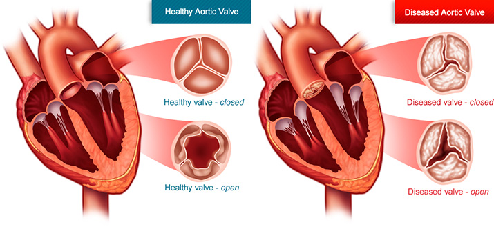
Clinical Assessment Of Aortic Stenosis
-
A. General Examination
(i) Blood pressure
- Old concept of systolic decapitation no longer holds true as severe aortic stenosis can be associated with normal systolic blood pressure in presence of associated aortic regurgitation, systemic hypertension and in elderly people.
2. Not only associated hypertension can be found in people with aortic stenosis, but that AS can itself cause systolic hypertension due to partial transmission of higher LV peak pressure across the aortic valve despite the stenotic valve acting as a pressure barrier. This may get completely corrected after AVR.
3. Significant aortic stenosis usually associated with a pulse pressure of 30 mm Hg or less
(ii) Arterial pulse
- Severe aortic stenosis is associated with typical slow rising pulse and late peaking pulse( parvus = small volume pulse, tardus = late peaking)
- This late rising and peaking of pulse in AS correlates with a gradient of 70 mm Hg or more across the aortic valve.
- In children with rapid velocity circulation and in elderly with non distensible arterial system, this sign may be absent.
- The late peaking component may not be appreciated in patients with heart failure/significant LV dysfunction where only low volume pulse may be appreciated
- Anacrotic pulse may also be found in patients with severe AS which is not palpable and usually a phonocardiogram event.
- Severe AS with heart failure/LV dysfunction may be associated with pulsus alternans.
- Factors that normalise arterial pulse and mask severity are
High cardiac output/elastic vessels in the young
Increased stiffness in elderly
Associated AR
Hypertension
8. Factors that exacerbate severity of AS
Decreased LV function
Hypovolemia
Associated mitral stenosis
9. Palpable thrill in carotid may be present.
10. Visible pulsations in the carotid due to turbulence across aortic valve(shudder) is observed in patients with aortic stenosis with associated aortic incompetence

(iii) JVP
- Prominent “a” wave with normal JVP—> due to LVH shifting the septum to right side and impaired RV filling—>Bernheim effect
2. If Biventricular failure—>mean JVP is raised
B. Cardiovascular Examination
(i) Precordial Motion
- Forceful LV lift with little or no displacement of point of maximal impulse(apex usually in 5th left ICS just shifted out and heaving apex in severe aortic stenosis)
2. Palpable A wave in supine/left lateral position(= S4) suggesting raised LVEDP or a thick non compliant chamber
3. Palpable S4 corresponds to a LV-Ao gradient of 70 mm Hg or more.
4. Features of pulmonary hypertension is usually not present in isolated aortic stenosis and if present should suspect associated mitral stenosis( other features being presence of short MDM, loud S1, AF, parasternal lift and opening snap).
5. A systolic thrill in right second ICS (may be radiating to carotid) best felt in sitting and leaning forward with breath held in expiration(also may be felt in suprasternal notch and right supraclavicular area(does not tell severity)
(ii) Heart Sounds
- S1 is usually unremarkable and may be decreased if
Increased LVEDP causing premature closure of mitral valve
associated LV dysfunction
Never accentuated so if loud indicates
Aortic ejection sound(very close to S1 and may get confused)
associated mitral stenosis
2. Second heart sound
(a) Intensity
Usually soft(calcific)
Normal/Accentuated in congenital AS with bicuspid valve
Older patients with aortic calcification or systemic hypertension it can be normal/loud
(b) Split
Can be narrow/single S2 due to increased LV ejection time
In severe AS, can be paradoxical split(splitting heard only in expiration and not during inspiration)
One must listen to splitting of S2 at apex,second RICS and second LICS away from maximum murmur intensity.
Causes of Paradoxical splitting
Delayed aortic valve closure Early pulmonic valve closure Severe Aortic stenosis WPW type B Severe AR RA myxoma(rare) HCM with obstruction TR(rare) Complete LBBB Severe LV dysfunction Acute MI During an episode of angina Severe hypertension RV pacing RV paced beats Patent ductus arteriosus(increased aortic hang out interval) 3. Third heart sound/fourth heart sound
S3 suggests LV dysfunction/CCF
S4 indicates a large LV-Ao gradient > 70 mm Hg and raised LVEDP
How does findings of AS change with LV dysfunction?
Soft S1 Pulsus alternans Apex shifted laterally and downward(sometimes diffuse apical impulse) Paradoxical split well appreciated LV S3 Only low volume pulse appreciated, tardus may not be appreciated Underestimation of aortic stenosis severity Late peaking component of murmur may not be well appreciated 4. Aortic Ejection Click
(a) Confirms organic nature of disease , localises anatomy to aortic valve and saysthat etiology is congenital bicuspid valve
(b) Related to maximal upward excursion of abnormal bicuspid valve
(c)Leaflet must be pliable. Uncommonly found in acquired AS in tricuspid valve
(d)high frequency, crisp sound occuring 40-80 msec after S1 and of snappy quality.
(e) Constant click (vs phasic of valvular PS, PS click best heard at base of heart)and best heard at apex and louder than S1/S2
(f) Loud discrete S1 heard at first and second ICS on either side of sternum raises possibility of ejection sound as loud S1 never heard at base of heart.
(g) A split at apex/LLSB may represent S1-ejection sound complex( click is a systolic event vs S4 a diastolic event; firm pressure with diaphgram or bell brings out a S1-click whereas attenuates S4)
(h) Causes of aortic vascular click
Systemic hypertension Anaemia/thyrotoxicosis Truncus arteriosus TOF Aneurysm of ascending aorta Aortic regurgitation 5. Murmur of Aortic stenosis
(a) Usually grade 4/6, medium pitch, mid systolic murmur, radiating to carotids and best heard in sitting and leaning forward with breath held in expiration with late peaking(signifies severe aortic stenosis)
(b)Gallavardin phenomenon – highest frequency components radiate to apex and may even sound musical at that site suggesting a murmur of MR(due to eddy currents)
(c) Factors where severity of AS underestimated and over estimated
Overestimated Underestimated Anaemia Heart failure/low CO/ LV dysfunction Thyrotoxicosis Polycythemia Pregnancy Associated proximal obstruction like MS, TS Associated AR Associated proximal regurgitation like MR, VSD Associated PDA Associated systemic hypertension/CoA(decreasing gradient) Tachycardia AF (d)Dynamic Auscultation
Inspiration decreases the murmur slightly(vs valvular PS) Expiration increased the murmur Increased on sitting and bending forward Standing decreases the murmur(vs HOCM) Valsalva decreases the murmur( decreases venous return) (vs HOCM) Squatting increases the murmur(increases venous return)(vs HOCM) Vasopressor increases murmur( increasing gradient, BP and contractility) Post PVC, murmurs increases(same as HOCM) Isometric hand grip decreases murmur by decreasing gradient(same as HOCM) (e) Differential diagnosis of AS murmur – SAVD, MR, HOCM
Aortic stenosis Sclerotic Aortic valve Long murmur with late peaking Murmur is shorter with no late peaking 4/6 less than 4/6 A2 diminished usually normal/increased Late rising arterial pulse usually normal S4 usually present absent Single A2/paradoxical split normal split LVH usually present usually absent Aortic stenosis Mitral regurgitation Usually radiate to carotid Radiate to axilla carotid thrill normal carotids parvus et tardus mini collapsing pulse Post PVC increase in murmur no beat to beat change, post PVC potentiation Amyl nitrate increases murmur decreases murmur Phenylephrine decreases murmur increases murmur Aortic stenosis HOCM Carotid pulse upstroke delayed and volume reduced Jerky pulse murmur well heard at base with radiation to apex LLSB with no radiation Usually associated with thrill usually no thrill S4 suggest severity S4 is pathognomic may have ejection click no click murmur decreases on valsalva murmur increases on valsalva murmur decreases on standing murmur increases on standing squatting increases murmur squatting decreases murmur C. Differentiating valvular, Sub valvular and Supra valvular obstruction
Valvular Sub valvular Supra valvular – – Elfin facies Absent Family Hx may be present Family Hx may be present – – Infantile hypercalcemia/renal failure Aortic ejection sound usually present if bicuspid Absent Absent Aortic Second heart sound usually soft Usually normal Normal Arterial pulse delayed upstroke with small amplitude Arterial pulse brisk upstroke with sometimes bisferiens Arterial pulse rapid upstroke in right carotid and delayed in left Absent Absent Right arm BP higher than left arm(Coanda effect) AR murmur can occur Congenital – almost all
Acquired – rareuncommon Murmur best in right second ICS and radiation to carotid Murmur best in left third/fourth ICS and no radiation Murmur best in right first ICS and no radiation to carotid Thrill usually present Absent Absent Aortic valve calcification can occur Absent Absent Ascending aortic dilatation usual Absent Absent Gradient is LV—> Ao LV—>LV Ao—>Ao (D)Features raising suspicion of aortic stenosis in patient with mitral valve disease
Angina or syncope Delayed carotid upstroke Prolonged ejection murmur LVH in presence of mitral stenosis on ECG Aortic valve calcium or post stenotic dilatation on CXR (E) Features raising suspicion of mitral stenosis in a patient with known aortic valve disease
Cough, dyspnea, orthopnea, PND, hemoptysis and sytemic emboli Parasternal lift Loud S1 Opening snap Diastolic murmur Presence of RVH on ECG Prominent pulmonary vascular markings on CXR Right ventricular dilatation on CXR (F) Clinical Assessors of severity in Aortic Stenosis
Pulsus parvus et tardus Paradoxical split Late peaking murmur LV S4 Apico-carotid delay Assessment of mixed valvular heart disease and multi-valvular heart disease shall be discussed later.


