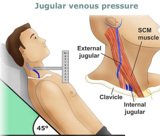
JVP in Congenital Heart Diseases
A. Acyanotic Congenital Heart Disease
- Atrial Septal Defect
(i). Small ASD’s usually do not have much effect on JVP.
(ii). In Large ASD’s(non-restrictive), “a” and “v” waves are equal. As age advances “a” wave becomes more prominent as LV compliance decreases. “a” wave becomes prominent with onset of pulmonary vascular disease and associated pulmonary stenosis.
(iii). Mean JVP may be raised in elderly indication RV/LV failure, lutembachers syndrome and underlying TAPVC.
(iv) In ostium primum ASD, JVP may have a prominent “v” wave(left atrial v wave) due to mitral regurgitation.
(v)In Lutembachers syndrome(ASD with rheumatic mitral stenosis), there is elevated JVP in the absence of right heart failure and elevated “a” wave in absence of PAH as right and left atrium act as a common chamber in non-restrictive ASD.
(vi)In AV septal defects, “v” wave is prominant as right atrium receives LV flow across an incompetent left AV valve directly through AV septal defect or indirectly through an ostium primum ASD.
2. Ventricular septal defect and PDA
(i) In post tricuspid shunts with moderately restrictive and non-restrictive defects with CCF, there is elevated mean JVP with prominent “a” and “v” waves.
(ii)In severe PS with restrictive VSD(pink TOF), a waves may be prominent.
(ii) Gerbode’s defect (LV to RA shunt) – As RA volume and distensibility increase in proportion to shunt, “V” waves though present are seldomly large even though LV ejects into RA.
3. Pulmonary Stenosis
(i) “a” waves are prominent as increases progressively as stenosis increases.
(ii)In PS , once RV failure sets in “v” waves becomes prominent.
(iii) Triology of fallot(PS with inter-atrial communication), hypertrophied RV causes right atrium to contract with greator force causing prominent “a” waves.
4. Congenitally corrected transposition of Great arteries
(i) As CC-TGA is associated with complete heart block, irregular slow cannon waves can be seen
(ii) Pulmonary stenosis can be associated with raised “a” wave amplitude which increases with severity
(iii)Pulmonary stenosis with non-restrictive atrial septal defect usually associated with normal JVP.
B. Cyanotic Congenital Heart Disease
- Tetralogy of Fallot
(i)JVP is normal in height and waveform as resistanve to right ventricle is at systemic and not supra-systemic level because of non restrictive VSD causing RV to decompress into aorta and also as right atrium is not required to increase its contractile force.
(ii) However “a” waves can be prominent in TOF in cases such as
Accessory tricuspid leaflet occluding the VSD
Systemic hypertension
Acquired stenosis of biventricular aortic valve
Adult TOF
(iii) Causes of elevated JVP in TOF includes anaemia, systemic hypertension, infective endocarditis(causing TR and AR), aortic regurgitation, broncho-pulmonary collaterals, adult TOF. associated PDA and after systemic to pulmonary shunt.
(iv)Persistent left superior vena cava may be associated with TOF which causes left sided jugular venous pulse elevation.
(iv) When RV fails in adult TOF, it produces TR and elevated “v” waves in JVP.
(vi) In TOF with absent pulmonary valve, JVP is elevated in parallel with elevated right ventricular filling pressure and “a” waves remain dominant unless RV failure sets in and “v” waves become prominent.
(vii) Similarly pulmonary atresia with intact ventricular septum produces prominent “a” waves in JVP.
2. Tricupid atresia
(i)”a ” waves are prominent when restrictive ASD obstructs egress blood flow from right atrium and also”y” descent is slow.”
(ii)”a” and “v” waves are bpoth increased when left ventricular failure causes a mean rise in left atrial pressure that is in continuity with right atrium.
(iii)Large “v” waves of mitral regurgitation are transmitted into right atrium when inter-atrial communication is non-restrictive.
3. Double outlet right ventricle(DORV)
(i) With sub-aortic DORV without PS, with increased pulmonary blood flow resulting in bi-ventricular failure, “a” “v” waves and mean JVP are elevated which normalizes when pulmonary vascular resistance increases. Similar applies to DORV with sub-pulmonic VSD(Taussig bing anomaly)
(ii) With sub-aortic VSD with pulmonary stenosis, “a” wave is normal because right ventricle ejects at but not above systemic vascular resistance similar to TOF.
4. Ebsteins Anomaly
(i) JVP is grossly normal( normal a and v waves and normal x descent despite severe tricuspid regurgitation) except “C” waves because of large mobile anterior tricuspid leaflet.
(ii) However. “a” waves can be prominent with a stenotic or imperforate tricuspid orifice
(iii) X descent and V waves are not prominent as (a)damping effect of roomy right atrium and thin walled toneless atrialised RV and (b) tricuspid regurgitation is low pressure and hypokinetic.
(iv) RV failure causes prominent “a” and “v” waves.
5. Complete Transposition of great arteries
JVP is elevated only in two conditions-
(i) Heart failure that accompanies a non restrictive VSD with low pulmonary vascular resistance(free communication)
(ii) RV failure in deeply cyanotic patients with a large volume of unsaturated blood recirculating through right ventricle and right atrium. (No communication)
6. Eisenmenger syndrome
(i)Only pre-tricuspid shunt like atrial septal defect can have supra-systemic pressures and hence in eisenmenger if “a” waves are prominent it must be ASD
(ii)In VSD, RV pressures can never exceed LV pressure and so “a” waves are never seen.
(iii) In PDA, rarely “a” waves can be present as theoretically supra-systemic pressures can occur and RV is decompressed by reversing ductal flow.
7. Single Ventricle
(i) Normally, height and waveforms are normal when moderate pulmonary stenosis curtails excessive pulmonary blood flow
(ii) Incompetence of right atrio-ventricular valve causes prominent “v” waves
(iii) Atresia of right atrio-ventricular valve increases “a” waves if inter-atrial communication is restrictive
(iv)Atresia of left atrio-ventricular valves causes large “a” waves provided atrial communication is non-restrictive.
In a nut shell,
(i) Though second heart sound is the most important factor in determining the diagnosis in congenital heart disease, jugular venous pulse also provides a helping aid.
(ii) In acyanotic shunt lesions, unless when pulmonary vascular disease sets in or RV failure develops, JVP doesn’t provide much aid to diagnosis
(iii)Consider the following conditions when there is prominent a wave in cyanotic congenital heart disease-
Tricuspid atresia
Pulmonary stenosis with intact ventricular septum and right to left shunt
TAPVC
Pulmonary atresia with intact ventricular septum
Adult TOF
TOF with restrictive VSD , systemic hypertension
(iv) Prominent “a” waves in JVP in eisenmenger syndrome suggests supra-systemic RV pressures and points inconspicuously towards ASD.