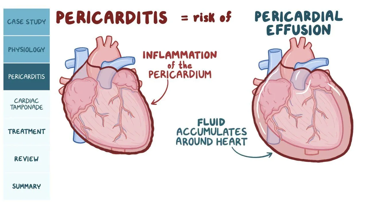
Pericardial Diseases and Management: Approach To Constrictive Pericarditis
A. CAUSES
idiopathic or viral etiology (42%-61%) |
postcardiac surgery (11%-37%) |
postradiation therapy primarily in Hodgkin’s lymphoma and breast cancer (2%-31%) |
connective tissue disease (3%-7%) |
postinfection, such as tuberculosis pericarditis (3%- 15% |
malignancy |
uremic pericarditis |
asbestosis |
trauma |
drug induced |
Idiopathic CP has the best prognosis followed by postsurgery CP with the worst prognosis in postradiation CP
Symptoms of right heart failure such as ascites, peripheral edema, and jugular venous pressure elevation with a rapid “y” descent.
Kussmaul’s sign was initially described for CP but can happen in any patients with increased right atrial pressure.
Dyspnea and pleural effusion are also presenting symptoms. Rarely, CP presents with severe hypoxemia caused by right to left shunt in patients with concomitant patent foramen ovale
B. PATHOPHYSIOLOGY

C. Diagnosis of CP
- ECHOCARDIOGRAPHY

2. CARDIAC CATHETERISATION


3. CXR/CT/MRI
Chest x-ray can detect pericardial calcifications especially on the lateral view
CT better evaluates pericardial anatomy (thickness/calcifications/effusion), as well as deformation of the cardiac contour
CT angiography allows for assessment of extent of pericardial calcification, its proximity to major coronary arteries, and areas of invasion into the myocardium.
In patients with prior cardiac surgery, CT defines the position of bypass grafts, and proximity of the right ventricle (RV) and great vessels to the sternum for safe redo sternotomy
Pericardial late gadolinium enhancement (LGE) on magnitude and phasesensitive inversion recovery sequences with and without fat suppression evaluates for pericardial vascularity, which is correlated with inflammation
Fat suppression helps distinguish abnormal pericardium with increased signal from epicardial fat.
T2 short tau inversion recovery imaging is performed to assess for pericardial edema, which is indicative of inflammation.
Positron emission tomography has been reported as a possible diagnostic tool for pericardial inflammation and can predict response to steroids in CP predominantly in patients with tuberculous pericarditis
D. TYPES OF CP

RECURRENT PERICARDITIS
RP is defined as the recurrence of pericarditis after a prior episode with at least 4 to 6 weeks of symptom-free interval, while incessant pericarditis usually lasts >4 to 6 weeks but
Etiologies include idiopathic, viral, bacterial (especially tuberculosis in developing world), postcardiac injury syndrome (eg, postpericardiotomy syndrome, post– transcatheter-based procedures, and postmyocardial infarction), postradiation, autoimmune disease (eg, systemic lupus erythematosus and rheumatoid arthritis), autoinflammatory, neoplastic (breast cancer, lung cancer, and lymphoma), uremia, and druginduced (eg, hydralazine)
The mean duration of RP that is difficult to treat ranges from 4.7 to 6.2 years from the onset time until complete therapy.
Current recommendations on the management of RP include NSAIDs and colchicine after initial relapse. Corticosteroids are used in patients who have refractory symptoms or cannot take NSAIDs.
The AIRTRIP (The Anakinra-Treatment of Recurrent Idiopathic Pericarditis) and RHAPSODY (Rilonacept Inhibition of Interleukin-1 Alpha and Beta for Recurrent Pericarditis: a Pivotal Symptomatology and Outcomes Study) trials demonstrated that IL-1 inhibitors (eg, rilonacept or anakinra) were an effective targeted therapeutic option for patients with colchicine-resistant and corticosteroid-dependent RP
Pericardiectomy can be considered in patients with RP refractory to medical therapy, dependent on corticosteroids, or intolerant to medical therapy.
E. DRUG MANAGEMENT

F. RISK STRATIFICATION

G. INDICATIONS FOR PERICARDIECTOMY

OUTCOMES OF PERICARDIECTOMY
In-hospital mortality of 6%
The overall 5-, 10-, and 20- year survival was 87%, 73% and 30%, respectively
Patients with an idiopathic etiology have the best outcomes with survival rates of >80% at 5 to 7 years with an operative mortality <1.5%
Postsurgery CP has survival 50% to 66% at 5 to 7 years of follow-up in several series with operative mortality of 4.5%
CP following mediastinal radiation was associated with 10.1% operative mortality and with 53.4% and 32.1% survival at 5 and 10 years, respectively
Patients with mediastinal radiation history have the worst short- and long-term outcomes.
PATIENTS WITH POOR CANDIDACY FOR PERICARDIECTOMY.
- Patients with “end-stage” CP.
- Patients with mixed CP with severe RCM
- Patients with end-stage renal disease and/or advanced liver disease with Child-Pugh B or C (score ‡7) or MELD-XI score (13.7-30.6)
- Patients with NYHA functional class I-II symptoms
- Patients who are not surgical candidates because of medical comorbidities
OPERATIVE PITFALLS
- Bleeding
- Injury to epicardial structures
- EPICARDIAL INFLAMMATORY RIND
- Myocardial calcification
- PRIOR CARDIAC SURGERY Patients with prior cardiac surgery, especially with patent coronary bypass grafts, present a challenge for successful Mediastinal radiation. Prior mediastinal radiation, especially for Hodgkin’s disease, presents a number of technical challenges
- Mediastinal radiation. Prior mediastinal radiation, especially for Hodgkin’s disease, presents a number of technical challenges
- Tricuspid and mitral valve regurgitation. Mitral and tricuspid valve evaluation is critical
- Phrenic nerve injury
(Adapted from JACC State of Art Review 2024)


